Microscope spec sheet
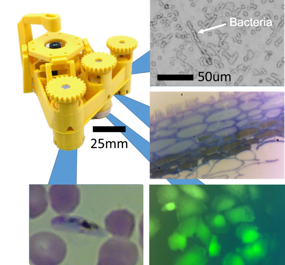
The Openflexure Microscope can be built in many different ways, from a low cost imager based on a $3 webcam to a lab-grade microscope complete with 100x oil immersion objective lens. This page gathers together some details on what's possible, though it's by no means an exhaustive list of how it can be configured.
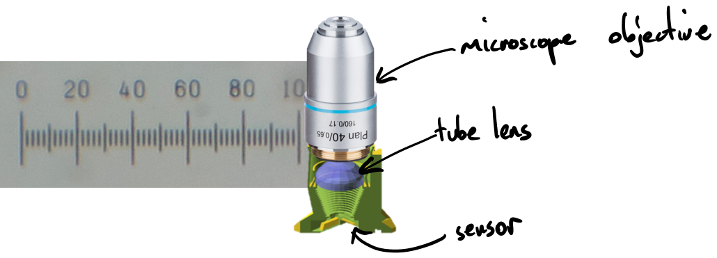
The simplest, cheapest set-up is to make a single-lens microscope, using the lens from the webcam you are using as an image sensor. This typically gets a resolution of better than a few microns; for the Raspberry Pi camera module version 2, the microscope can resolve down to 1-2um with a field of view of 280x210um. For higher-end work, an RMS microscope objective can be used, together with a tube length correction lens that increases the field of view for small camera sensors. The highest magnification configuration that's commonly used is a 100x oil immersion objective, which has a field of view around 141x106um and a resolution better than 500nm, around the diffraction-limited resolution set by the objective.
The basic microscope is configured to produce images in bright-field transmission mode, using either a single LED or a single-lens condenser assembly. However, other configurations are possible and are gradually being documented and included in the main design. These include epifluorescence (using a miniature filter cube and LED illumination) and dark field (using a custom condenser assembly).
A plastic flexure stage gives smooth control over focus and sample translation. It can be automated with three low-cost, low-power stepper motors, which have a step size of <100nm and a repeatability of around 1um. The range of travel is a little over 10x10x4mm for the LS65 version of the stage, and 7x7x4mm for the SS40 version. A detailed study of the mechanical performance is given in Review of Scientific Instruments (open access).